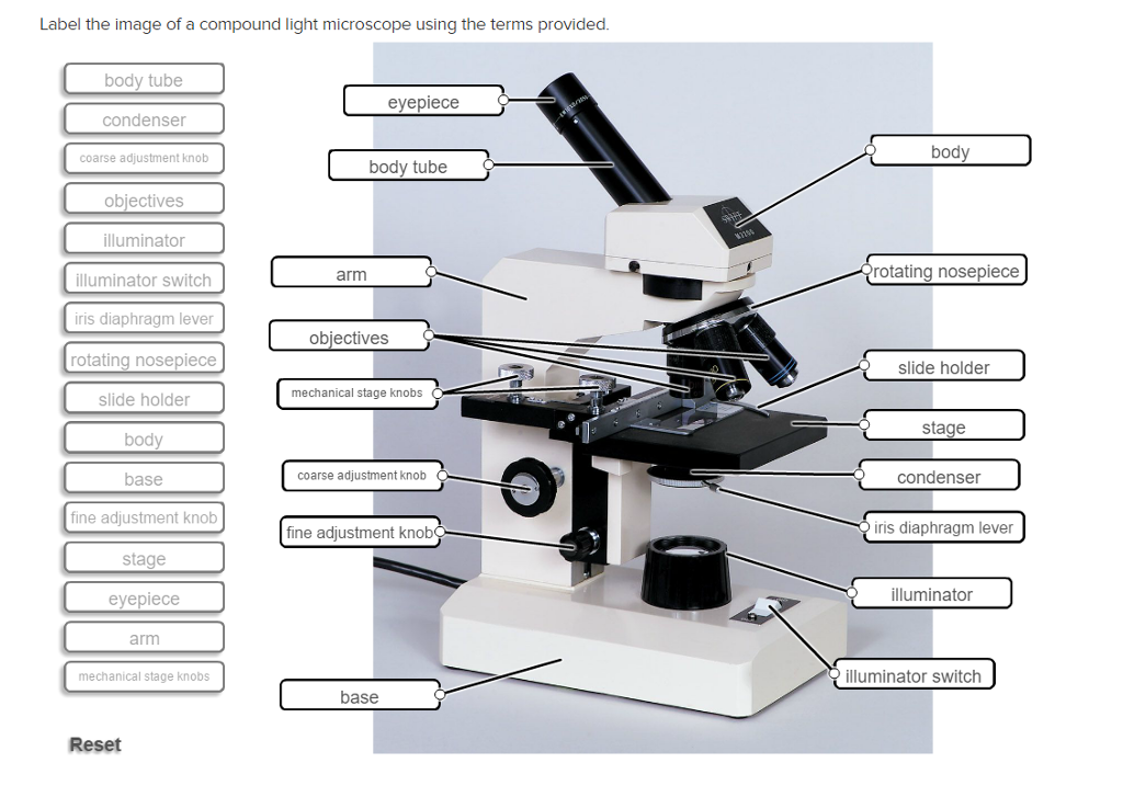
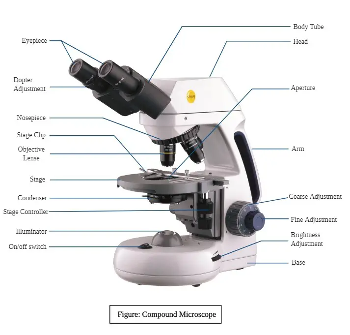
38 compound light microscope with label
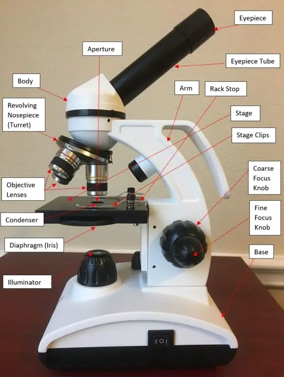
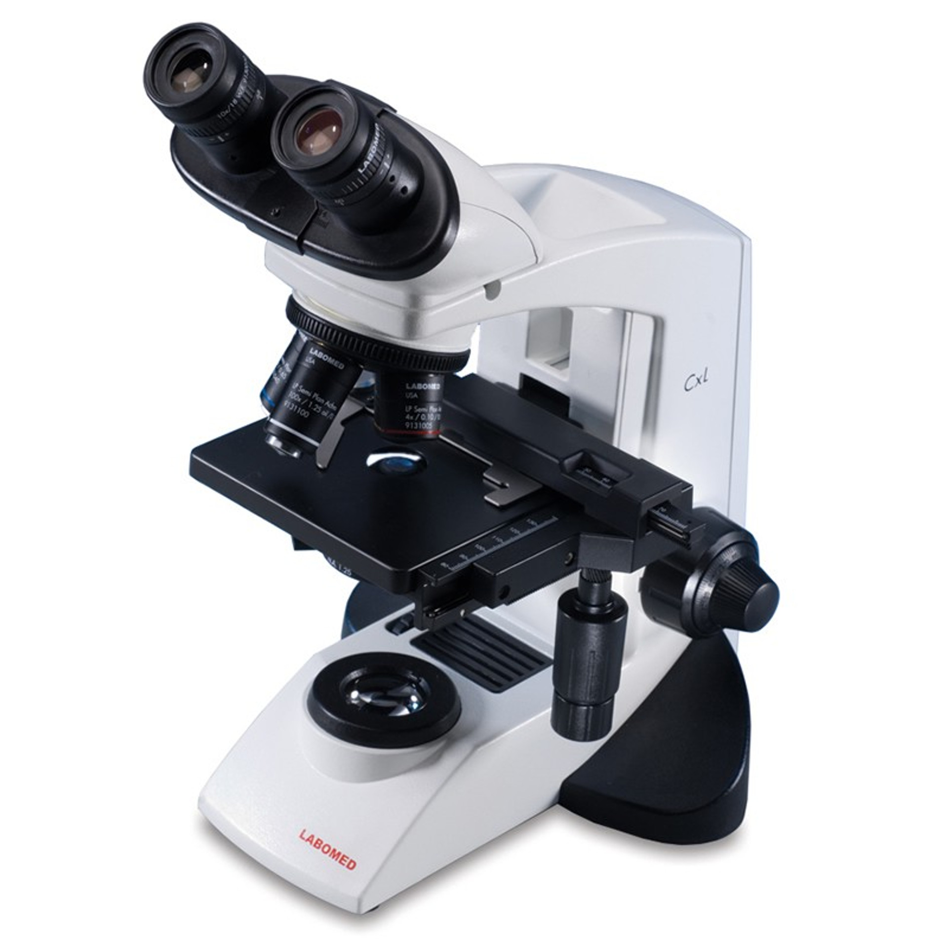
Compound Microscope Parts, Functions, and Labeled Diagram Compound Microscope Definitions for Labels Eyepiece (ocular lens) with or without Pointer: The part that is looked through at the top of the compound microscope. Eyepieces typically have a magnification between 5x & 30x. Monocular or Binocular Head: Structural support that holds & connects the eyepieces to the objective lenses. The Compound Light Microscope: Optics, Magnification and Best Used For 23 The Compound Light Microscope is an optical microscope that uses light to magnify objects. It is one of the most commonly used microscopes in scientific research and has been instrumental in furthering our understanding of the natural world. The compound light microscope consists of two lenses: the eyepiece lens and the objective lens.
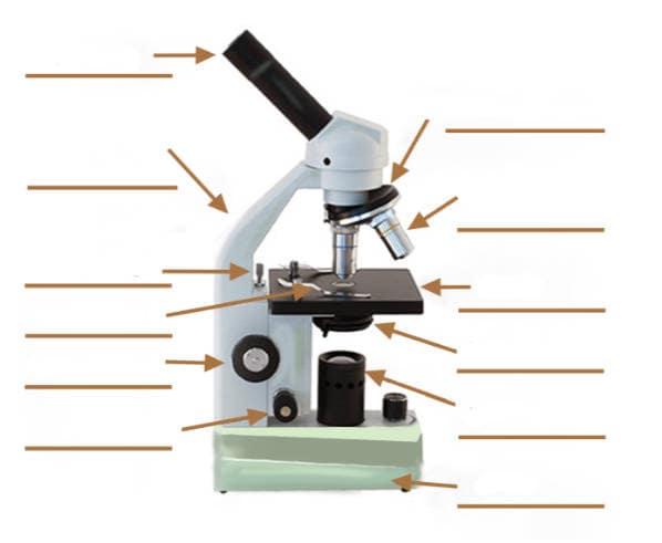
Labeling the Compound Light Microscope Game - PurposeGames.com Labeling the Compound Light Microscope Game — Quiz Information. This is an online quiz called Labeling the Compound Light Microscope Game. There is a printable worksheet available for download here so you can take the quiz with pen and paper.

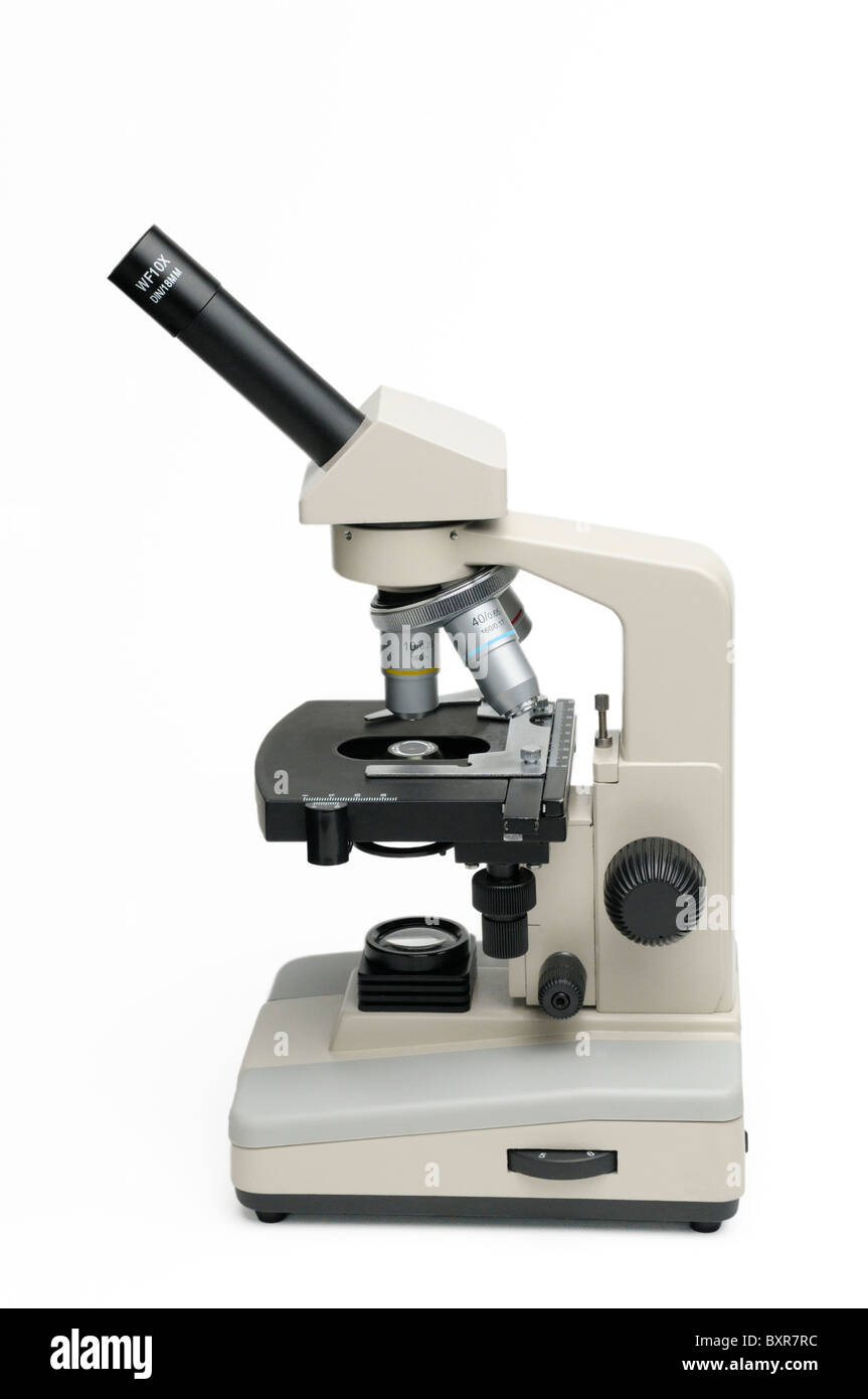
Compound light microscope with label
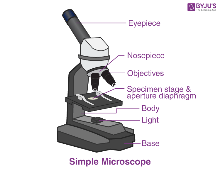
Compound Microscope- Definition, Labeled Diagram, Principle, Parts, Uses A compound light microscope is relatively small, therefore it's easy to use and simple to store, and it comes with its own light source. Because of their multiple lenses, compound light microscopes are able to reveal a great amount of detail in samples. Read Also: Light Microscope- Definition, Principle, Types, Parts, Labeled Diagram, Magnification Compound Light Microscope: Everything You Need to Know A compound light microscope is a type of light microscope that uses a compound lens system, meaning, it operates through two sets of lenses to magnify the image of a specimen. It's an upright microscope that produces a two-dimensional image and has a higher magnification than a stereoscopic microscope. It also goes by a couple of other names ... PDF Parts of the Light Microscope - Science Spot Supports the MICROSCOPE D. STAGE CLIPS HOLD the slide in place C. OBJECTIVE LENSES Magnification ranges from 10 X to 40 X F. LIGHT SOURCE Projects light UPWARDS through the diaphragm, the SPECIMEN, and the LENSES H. DIAPHRAGM Regulates the amount of LIGHT on the specimen E. STAGE Supports the SLIDE being viewed K. ARM Used to SUPPORT the
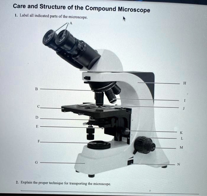
Compound light microscope with label. Identification of Parts and Functions: Label each | Chegg.com Expert Answer. Transcribed image text: Identification of Parts and Functions: Label each part of the compound light microscope and learn the function of each. Use information in the lab resources folder to help you. Note that the microscope below lacks a mechanical stage. It also had an iris diaphragm under the stage instead of a disk diaphragm. Compound Microscope - Diagram (Parts labelled), Principle and Uses Also called as binocular microscope or compound light microscope, it is a remarkable magnification tool that employs a combination of lenses to magnify the image of a sample that is not visible to the naked eye. Compound microscopes find most use in cases where the magnification required is of the higher order (40 - 1000x). Compound Microscope: Definition, Diagram, Parts, Uses, Working ... - BYJUS A compound microscope is defined as A microscope with a high resolution and uses two sets of lenses providing a 2-dimensional image of the sample. The term compound refers to the usage of more than one lens in the microscope. Also, the compound microscope is one of the types of optical microscopes. 2 Microscope Lab Worksheet.pdf - Using a Compound Light... Instructions for Lab: Log onto Canvas and complete the virtual activity on Using a Microscope. Download and read and take notes on the " Using a Compound Light Microscope " pdf. Watch the video linked in Part A: Video 1 and answer the questions about the video. Then label the pictures of the compound microscope in Part B, using information from both the simulation and the pdf.
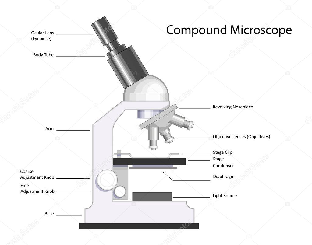
Light Microscope Labeled Diagram, Definition, Principle, Types, Parts ... Parts of a bright-field microscope or Compound light microscope. An optical microscope, the bright-field microscope (or compound light microscope) is an invaluable tool in the fields of biology, medicine, and education. ... (January 31, 2023).Light Microscope Labeled Diagram, Definition, Principle, Types, Parts, Applications. Retrieved from ... Binocular Microscope Anatomy - Parts and Functions with a Labeled ... The optical part of the compound light microscope includes - Condenser with iris diaphragm, Illuminator (light source), An objective lens of the microscope, and The eyepiece or ocular lens of the microscope So, the other parts of the microscope (except these four) are included under the non-optical components. Compound Microscope Labeled Diagram | Quizlet Compound Microscope Labeled + − Flashcards Learn Test Match Created by meganplocher734 Terms in this set (14) Eyepiece/Ocular lens Contains the ocular lens Body tube A hollow cylinder that holds the eyepiece. Arm Part that supports the microscope. Stage Supports the slide or specimen Coarse adjustment Knob Parts of the Microscope with Labeling (also Free Printouts) 5. Knobs (fine and coarse) By adjusting the knob, you can adjust the focus of the microscope. The majority of the microscope models today have the knobs mounted on the same part of the device. Image 5: The circled parts of the microscope are the fine and coarse adjustment knobs. Picture Source: bp.blogspot.com.
Compound Light Microscope Optics, Magnification and Uses Magnification. In order to ascertain the total magnification when viewing an image with a compound light microscope, take the power of the objective lens which is at 4x, 10x or 40x and multiply it by the power of the eyepiece which is typically 10x. Therefore, a 10x eyepiece used with a 40X objective lens, will produce a magnification of 400X. Parts of a Microscope with Their Functions • Microbe Online The common light microscope used in the laboratory is called a compound microscope. It is because it contains two types of lenses; ocular and objective. The ocular lens is the lens close to the eye, and the objective lens is the lens close to the object. These lenses work together to magnify the image of an object. Solved Label the image of a compound light microscope using - Chegg Expert Answer. 100% (17 ratings) Transcribed image text: Label the image of a compound light microscope using the terms provided. Parts of the Compound Microscope - Houston Community College Parts of the Compound Microscope Use Figure 2 as a guide to locate the major parts of the compound microscope. a. Base: The bottom, flat part that supports the microscope. b. Arm: The straight or curved vertical part that connects the base to the upper portion. c. Body Tube: Extends from the arm and contains the ocular lens and the rotating
1.5: Microscopy - Biology LibreTexts In Biology, the compound light microscope is a useful tool for studying small specimens that are not visible to the naked eye. The microscope uses bright light to illuminate through the specimen and provides an inverted image at high magnification and resolution. ... Blank microscope to label parts. This page titled 1.5: Microscopy is shared ...
Compound Microscope Parts The three basic, structural components of a compound microscope are the head, base and arm. Head/Body houses the optical parts in the upper part of the microscope Base of the microscope supports the microscope and houses the illuminator Arm connects to the base and supports the microscope head. It is also used to carry the microscope.
2.5: Use of Compound Light Microscopes for Anatomy Laboratories Focusing Procedure for a Compound Light Microscope. Place the microscope on a table in front of you and plug it in such that the cord does not create a trip hazard for you or others in the laboratory. Turn the revolving nosepiece to have lowest magnification objective in place, usually 4x. The objective should click when properly in place.
Compound Light Microscope Labelling Quiz - PurposeGames.com Compound Light Microscope Labelling by LearnAnatomy 2,246 plays 15 questions ~40 sec English 4 too few (you: not rated) Tries 15 [?] Last Played February 22, 2022 - 12:00 am There is a printable worksheet available for download here so you can take the quiz with pen and paper. Remaining 0 Correct 0 Wrong 0 Press play! 0% 08:00.0 Highscores
Label Parts of a Compound Light Microscope Flashcards | Quizlet Study with Quizlet and memorize flashcards containing terms like Arm, Base, Diaphragm and more.
Microscope Parts and Functions Here are the important compound microscope parts... Eyepiece: The lens the viewer looks through to see the specimen. The eyepiece usually contains a 10X or 15X power lens. Diopter Adjustment: Useful as a means to change focus on one eyepiece so as to correct for any difference in vision between your two eyes.
Label the microscope — Science Learning Hub Label the microscope Interactive Add to collection Use this interactive to identify and label the main parts of a microscope. Drag and drop the text labels onto the microscope diagram. eye piece lens diaphragm or iris coarse focus adjustment stage base fine focus adjustment light source high-power objective Download Exercise Tweet
Parts of the Microscope (Labeled Diagrams) - Simple and Compound Microscope A compound microscope is a complicated assembly of various lenses that produces a significantly enlarged image of minute living things as well as other delicate characteristics of cells and tissues. The parts of the compound microscope is categorized into two - the mechanical parts and the optical parts.
Parts of a microscope with functions and labeled diagram - Microbe Notes Microscopic illuminator - This is the microscopes light source, located at the base. It is used instead of a mirror. It captures light from an external source of a low voltage of about 100v. Condenser - These are lenses that are used to collect and focus light from the illuminator into the specimen.
Compound Light Microscope labeled Functions Best Uses, Parts 23 A compound light microscope labeled is a type of light microscope that uses two or more lenses to magnify an object. The first lens, called the objective lens, is positioned near the object being viewed. A second lens, called the eyepiece, is located near the viewer's eye.
PDF Parts of the Light Microscope - Science Spot Supports the MICROSCOPE D. STAGE CLIPS HOLD the slide in place C. OBJECTIVE LENSES Magnification ranges from 10 X to 40 X F. LIGHT SOURCE Projects light UPWARDS through the diaphragm, the SPECIMEN, and the LENSES H. DIAPHRAGM Regulates the amount of LIGHT on the specimen E. STAGE Supports the SLIDE being viewed K. ARM Used to SUPPORT the
Compound Light Microscope: Everything You Need to Know A compound light microscope is a type of light microscope that uses a compound lens system, meaning, it operates through two sets of lenses to magnify the image of a specimen. It's an upright microscope that produces a two-dimensional image and has a higher magnification than a stereoscopic microscope. It also goes by a couple of other names ...
Compound Microscope- Definition, Labeled Diagram, Principle, Parts, Uses A compound light microscope is relatively small, therefore it's easy to use and simple to store, and it comes with its own light source. Because of their multiple lenses, compound light microscopes are able to reveal a great amount of detail in samples. Read Also: Light Microscope- Definition, Principle, Types, Parts, Labeled Diagram, Magnification














Komentar
Posting Komentar