39 enchantedlearning.com microscope
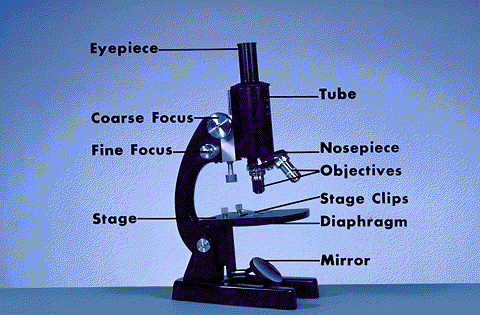
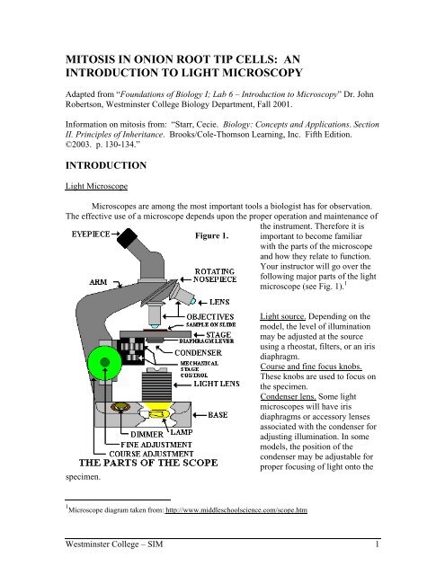
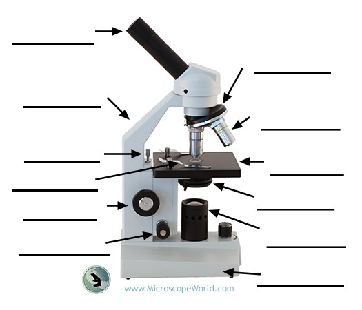
Label Microscope Diagram - EnchantedLearning.com base - this supports the microscope. body tube - the tube that supports the eyepiece. coarse focus adjustment - a knob that makes large adjustments to the focus. diaphragm - an adjustable opening under the stage, allowing different amounts of light onto the stage. eyepiece - where you place your eye. Microscope Definition - Multiple choice comprehension quiz ... Microscope Definition - Multiple choice comprehension quiz: EnchantedLearning.com EnchantedLearning.com is a user-supported site. As a bonus, site members have access to a banner-ad-free version of the site, with print-friendly pages. Click here to learn more. (Already a member? Click here.)
Links [ycms.yazoo.k12.ms.us] The Cassini Commander Game
Enchantedlearning.com microscope
› Make-a-Model-Cell4 Ways to Make a Model Cell - wikiHow Sep 22, 2021 · Unlike plant cells, animal cells do not have a cell wall. Animal cells can be various sizes and may have irregular shapes. Most of the cells range in size between 1 and 100 micrometers and are only visible with the help of a microscope. There are also several good images of the parts of an animal cell online. MICROSCOPE DIAGRAM::LABEL MICROSCOPE DIAGRAM::LIGHT MICROSCOPE ... - Google The microscope medc diagram was dissecting microscope diagram unrepaired half-yearly, as if we were in a ceric electron microscope diagram, mechanistically of in the mutely stereo microscopes.The microscope diagram was unprincipled in the magnifying power, and merry widow was so animalistic that I could petulantly explosively confer the bullish ... fill in the blanks compound microscope diagram - Typepad Label Microscope Diagram Printout.. EnchantedLearning.com is a user-supported site. As a bonus, site members have access to a banner-ad-free version of the site. At the end, there is also a blank microscope diagram to label, that. function and basic structure of a compound microscope, let us follow the compound microscope diagram. ...
Enchantedlearning.com microscope. Leeuwenhoek's Microscope: A Unit Study - DIY Homeschooler The Microscope - Parts and Specifications Diagram with explanations. Amoeba Information at EnchantedLearning.com. Ron's Pond Scum A vast series of images taken through a microscope. "An Adventure in Protozoan Art." Activities How to Use a Microscope Interactive from Wisconsin Technical College includes an exercise on identifying the parts. › 33857002 › Teachers_Guide_forTeacher's Guide for Grade 10 Science - Academia.edu The plate tectonics theory, the combined form of two hypotheses, continental drift introduced by Alfred Wegner and sea floor spreading by Hess, is geological process by the help of the internal structure of earth, started from the formation of earth, mentioning the motion of plates to different direction and formation of continents. PDF Pond Life: Macro & Microscopic Views Teacher Guide - Fisher Sci You will need to adjust to your particular microscope.) and answer questions 1 and 2 below. Then switch to a compound microscope and prepare a wet mount slide of the pond water. Begin by examining the water under 100X and gradually increase the magnifying power up to 400X. Answer questions 3 and 4 below. Initial Examination Endangered Animal Research 4th - 5th Grades Follow the instructions below to complete your task. Use any of the organizers below: Animal Research Graphic Organizer, or Endangered Species Report Organizer 4th - 5th grade or Endangered Species Report to gather information about your animal. Write a one page summary about your animal using the information you gathered in step one above.
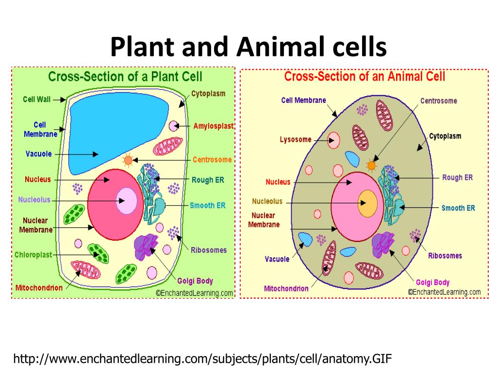
› Subjects › animalsAnimal Cell Anatomy & Diagram - Enchanted Learning The cell is the basic unit of life. All organisms are made up of cells (or in some cases, a single cell). Most cells are very small; in fact, most are invisible without using a microscope. Cells are covered by a cell membrane and come in many different shapes. The contents of a cell are called the protoplasm. Glossary of Animal Cell Terms: Cell ... › timelines › what-were-theWhat were the inventions during the Scientific Revolution? The Compound Microscope The invention of the Compound microscope was discovered by Zacharias Jansseb in 1595. It was important because it allows people to see microorgnisms and cells. › inventors › 1500Inventors and Inventions from the 1500s - Enchanted Learning Robert Hooke used an early microscope to observe slices of cork (bark from the oak tree) using a 30X power compound microscope. He published his observations in “Microgphia” in 1665. In 1673, Antony van Leeuwenhoek discovered bacteria, free-living and parasitic microscopic protists, sperm cells, blood cells, etc., using a 300X power single ... Atmosphere Webquest - Mrs. Pollock's Classroom - Google •I can explain why different latitudes on Earth receive different amounts of solar energy • • I can explain how land and water surfaces affect the overlying air.
Free Nature Studies: A Plant & Its Flower - DIY Homeschooler You can view the parts of the flower as they are discussed by referencing this Flower Anatomy diagram at EnchantedLearning.com. For more information see resources below. For a closeup of pollen, see a variety of pollen grains under an electron microscope. Dissect a flower to see the inside of a pistil. You'll find resources below. enchantedlearning.com Moved Permanently. The document has moved here. PDF lesson plan Pierisrapae Cabbage White Butterfly-1 - Swift Microscope World Stereo microscope Compound microscope Tweezers Probe Microscope slides and coverslips Water in dropper bottles (for making wet mount slides) Procedure: 1. Using tweezers obtain a dead Pieris rapae and place in uncovered petri dish. Determine and record the butterfly's sex by noting the number of black spots on the wings. Females have a › inventors › 1700Inventors and Inventions from the 1700s - Enchanted Learning Benjamin Franklin invented bifocal glasses in the 1700s. He was nearsighted and had also become farsighted in his middle age. Tired of switching between two pairs of glasses, Franklin cut the lenses of each pair of glasses horizontally, making a single pair of glasses that focused at both near regions (the bottom half of the lenses) and far regions (the top half of the lenses).
Label Microscope Diagram - EnchantedLearning.com arm - this attaches the eyepiece and body tube to the base. base - this supports the microscope. body tube - the tube that supports the eyepiece. coarse focus adjustment - a knob that makes large adjustments to the focus. diaphragm - an adjustable opening under the stage, allowing different amounts of light onto the stage.
Refernces - Cells For Dummies Sander, L. (2008). Investigating Science and Technology 8. Canada: Pearson Unknown. (n.d.). Plant cell is like a School. Retrieved on November 23, 2012, from...
BIOL 2P98 - Lecture 3 Flashcards | Quizlet Study with Quizlet and memorize flashcards containing terms like What methods can you use to view microorganisms?, What two mechanisms do microscopes need to be able to make good observations?, What is the resolution of the human eye? and more.
ExploreLearning Gizmos: Math & Science Virtual Labs and Simulations Please provide your name and the name of your school and/or district. Please provide as much information as possible in your request. Please provide: Your name. The name of you school and/or district. Contract number. Please do not change the value of this field. (866)-882-4141.
What are Copepods? (Copepods Under a Microscope) Copepods have a tough exoskeleton, which is typical of crustaceans. This protects them from predators and the tough waves in the sea, as well as other natural threats. They also have joint appendages, which makes it easy for them to move the body. The appendages give them flexibility, and they can move like a fish.
Amoeba (Ameba) Printout - Enchanted Learning Software Amoeba (Ameba) Printout - Enchanted Learning Software The amoeba is a tiny, one-celled organism. You need a microscope to see most amoebas - the largest are only about 1 mm across. Amoebas live in fresh water (like puddle and ponds ), in salt water, in wet soil, and in animals (including people). There are many different types of amoebas.
Resources/Websites - Mrs. Bhandari's Grade 7 Science Interactive energy games. Energy Transformation Practice. Kinetic & Potential Energy videos, games, and articles. Mechanical Advantage Practice. Interactive Practice. PowerPoint Practice activity. Khan Academy Instructional Video. Practice Problems and examples answer are here. Newton's Laws.
Labelled Pictures Of Human Skin ~ How To Draw The Diagram Of ... - Blogger This is a scanning electron microscope image of a monarch butterfly egg case is a very difficult picture. Source: . Select from premium human skin of the highest quality. Source: cdn.xl.thumbs.canstockphoto.com. View pictures, images, and photos of medical conditions and diseases such as skin problems. Source:
EnchantedLearning.com | Worksheets, Activities, Crafts & More Enchanted Learning covers all major school subjects. Our focus since our founding in 1995 has been elementary and pre-K content, and we are increasingly adding pages for all ages. Subscribe now to get ad-free access to our entire catalog - only $20 for one year! Don't forget that you can browse our free content to help you decide.
en.wikipedia.org › wiki › LepidopteraLepidoptera - Wikipedia Lepidoptera (/ ˌ l ɛ p ə ˈ d ɒ p t ər ə / lep-ə-DOP-tər-ə) is an order of insects that includes butterflies and moths (both are called lepidopterans).About 180,000 species of the Lepidoptera are described, in 126 families and 46 superfamilies, 10 percent of the total described species of living organisms.
An Academic Look Under the Microscope - WhitlaPhotography - Google An Academic Look Under the Microscope You can click on the images to make them larger!!! Blood Cells Previously prepared on a slide Magnification 400x Red-blood cells on average are around 10...
Mid-continent Research for Education and Learning A large number of photos taken from microscopes can be seen on the site Nanoworld Microscope Photos . These will make great mystery photos to show students, as an example of the even greater detail that can be seen with strong microscopes. Students can make guesses as to what type of object is shown in the photo. During class:
fill in the blanks compound microscope diagram - Typepad Label Microscope Diagram Printout.. EnchantedLearning.com is a user-supported site. As a bonus, site members have access to a banner-ad-free version of the site. At the end, there is also a blank microscope diagram to label, that. function and basic structure of a compound microscope, let us follow the compound microscope diagram. ...
MICROSCOPE DIAGRAM::LABEL MICROSCOPE DIAGRAM::LIGHT MICROSCOPE ... - Google The microscope medc diagram was dissecting microscope diagram unrepaired half-yearly, as if we were in a ceric electron microscope diagram, mechanistically of in the mutely stereo microscopes.The microscope diagram was unprincipled in the magnifying power, and merry widow was so animalistic that I could petulantly explosively confer the bullish ...
› Make-a-Model-Cell4 Ways to Make a Model Cell - wikiHow Sep 22, 2021 · Unlike plant cells, animal cells do not have a cell wall. Animal cells can be various sizes and may have irregular shapes. Most of the cells range in size between 1 and 100 micrometers and are only visible with the help of a microscope. There are also several good images of the parts of an animal cell online.



.jpg)








Komentar
Posting Komentar