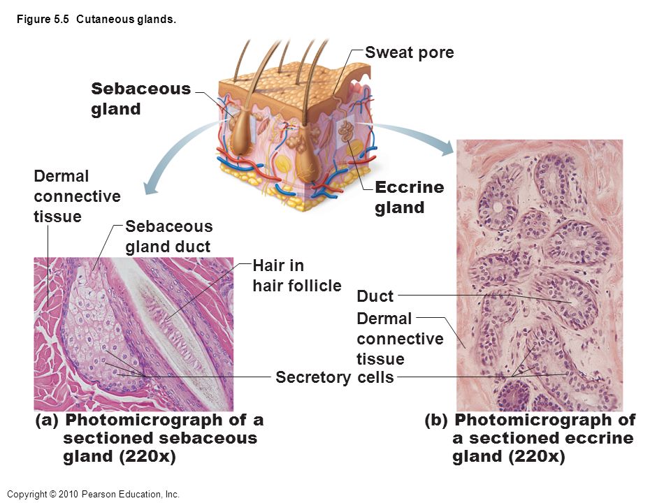
39 label the photomicrograph of the sebaceous gland.
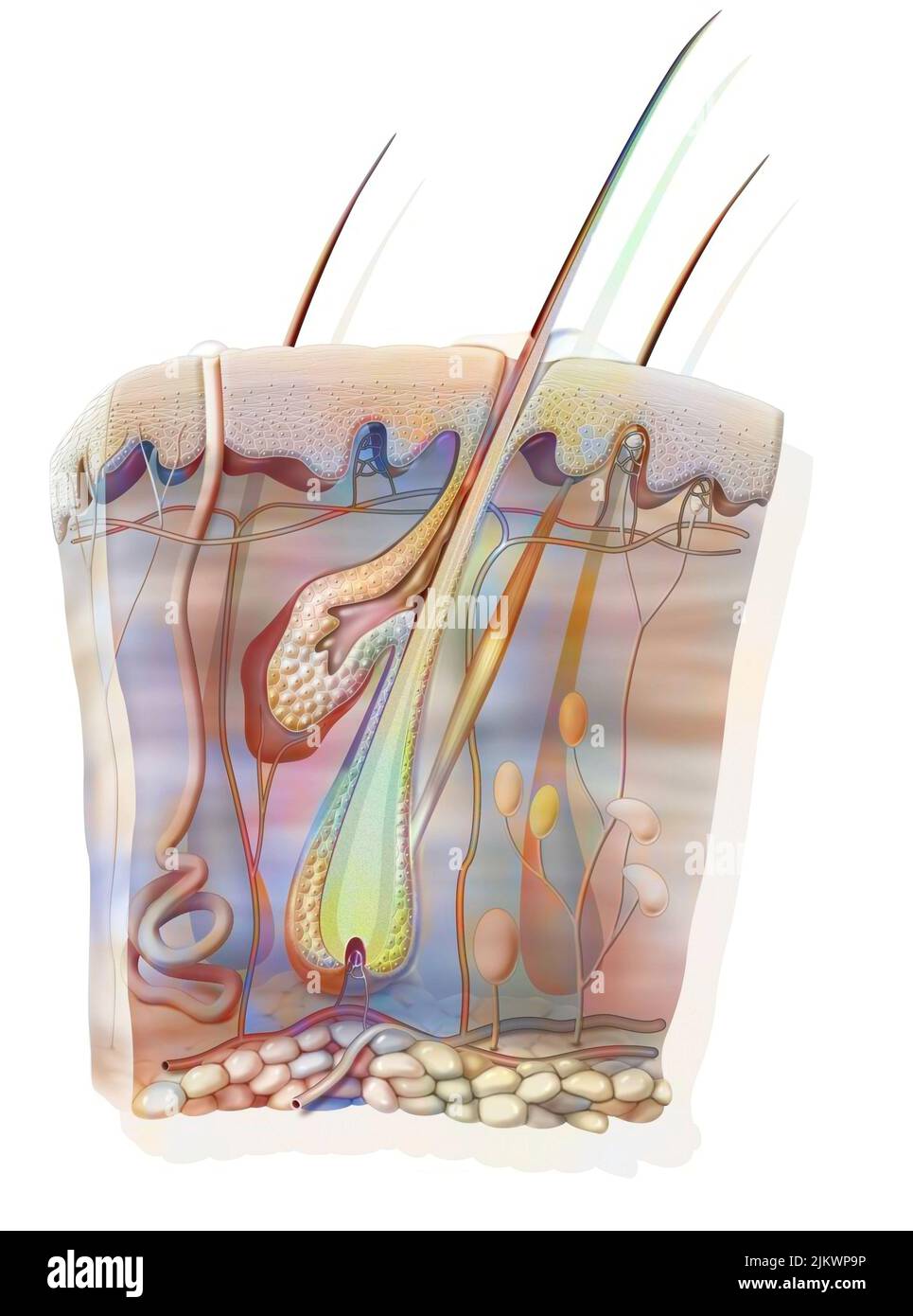
PDF Name the Condition the cartoon and the photomicrograph. Name the 4 layers of thin skin in both the cartoon and the photomicrograph. •Name the Layers of skin and label the dermal papilla and dermis •Name the Layers of skin and label the dermal papilla and dermis. Name the layer of skin shown. Stratum Spinosum. ... Sebaceous gland • Identify the following ... Anatomy and Physiology Homework Chapter 6 Flashcards Study with Quizlet and memorize flashcards containing terms like Label the parts of the skin and subcutaneous tissue. -Blood Capillaries -Piloerector muscle -Dermal papilla -Hair bulb -Sensory nerve fibers -Tactile corpuscle -Hair follicle -Sebaceous gland, Label the parts of the skin and subcutaneous tissue. -Hypodermis -Sweat pores -Dermis -Hairs -Cutaneous blood vessels …
Sebaceous Gland - an overview | ScienceDirect Topics Sebaceous glands are more numerous along the dorsum in dogs and sparse along the ventrum—an important factor to consider when sampling for sebaceous adenitis. ... Photomicrograph of Zymbal's gland tumor showing the typical mixed squamous (arrow) and sebaceous differentiation. The arrowhead depicts a nest of plump neoplastic sebaceous ...
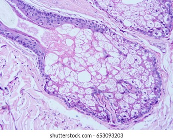
Label the photomicrograph of the sebaceous gland.
Answered: • hair bulbs • hair follicle • hair… | bartleby Solution for • hair bulbs • hair follicle • hair root • papilla of hair • sebaceous gland 1 1 2 2 3 3 4 5 4 5 10x Courtesy Michael Ross, University of Florida Pyridine and 4-dimethylaminopyridine (DMAP) are pictured...open 8 Label the photomicrograph of thin skin. Hair Sebaceous gland Dermis Hair Follicle Epidermis Duct of sebaceous gland KS Name the structure. Type here to search T Chapter 6 - Labeling Parts of Skin - sweat gland blood... Labeling Parts of the Skin Identify the layers of skin. dermis stratum basale stratum spinosum stratum lucidum stratum corneum stratum granulosum basement membrane Identify the parts of this photomicrograph of skin: sebaceous gland hair shaft hair follicle dermis epidermis End of preview. Want to read all 2 pages? Upload your study docs or become a
Label the photomicrograph of the sebaceous gland.. Quiz #3 Study Guide Flashcards | Quizlet Correctly label the following areas on a slide of areolar connective tissue. Label the tissues found in the stomach with the items provided. Terms will be used more than once. Match these cells found in connective tissues to their functions. 1. Cells that form fibers and ground substance in the extracellular matrix FIBROBLASTS 2. Sebaceous Gland Label The Photomicrograph Of Thin Skin - Blogger Sebaceous Gland Label The Photomicrograph Of Thin Skin - Integumentary System Histology Post a Comment And lymph vessels, nerves, and other structures, such as hair follicles and sweat glands. Using the slide thin skin with hairs, and the photomicrographs of cutaneous glands (figure 7.7) as . This problem has been solved! Show a separate graph of the constraint lines and the...open 8 Label the photomicrograph of thin skin. Hair Sebaceous gland Dermis Hair Follicle Epidermis Duct of sebaceous gland KS Name the structure. Type here to search T Quiz #3 Study Guide Flashcards | Quizlet sebaceous secretes sebum, holocrine gland that usually opens up into a hair follicle, coats the scalp hair with oil, blockage and infection ... Label the photomicrograph of thin skin. Organize the following layers of the epidermis from superficial to deep. Categorize the appropriate structures or descriptions with the appropriate layer of skin ...
Question : Label the photomicrograph of the sebaceous gland. - Chegg Expert Answer. 100% (30 ratings) Transcribed image text: Label the photomicrograph of the sebaceous gland. CH 5 Integument - CHAPTER 5 INTEGUMENT Skin (Integument)... - Course Hero Sebaceous (Oil) Glands • Widely distributed - Not in thick skin of palms and soles • Most develop from hair follicles and secrete into hair follicles • Relatively inactive until puberty - Stimulated by hormones, especially androgens • Secrete sebum - Oily holocrine secretion - Bactericidal - Softens hair and skin Bio Lab Chapter 6 Quiz Flashcards | Quizlet -hair follicle and sebaceous gland Identify the type of tissue that composes the epidermis of the skin. stratified squamous epithelial tissue Identify the structures of the dermis. dense connective tissue with fibers oriented in many directions dense irregular loose connective tissue characterized by long, thin dark fiber areolar tissue Kamus Kedokteran Dorland Edisi 31 [z0x292o9mwqn] [Yun.] lebih kecil. rriosis mikros fYrn.l kecil. Juga menunjukkan fraksi dalam sistem metrik (satu per seiuta). photomicrograph, microgram mille lL.l seribu. Juga menunjukkan fraksi dalam sistem metrik. Cf. kilo-. milligram, tnillipede mimetikos [Yun.] meniru. sympalhomimetic nisos [Yun.l kebencian. rzisogamy Lihat -mittent. intromlssion
Anatomy and Physiology Homework Chapter 6 Flashcards | Quizlet Study with Quizlet and memorize flashcards containing terms like Label the parts of the skin and subcutaneous tissue. -Blood Capillaries -Piloerector muscle -Dermal papilla -Hair bulb -Sensory nerve fibers -Tactile corpuscle -Hair follicle -Sebaceous gland, Label the parts of the skin and subcutaneous tissue. -Hypodermis -Sweat pores -Dermis -Hairs -Cutaneous blood vessels -Epidermis -Sweat ... Final Exam A&P 1 Flashcards | Quizlet Label the photomicrograph of thin skin Hair shaft, epidermis, dermal root sheath, sebaceous gland, dermis, hair matrix label the structures of the hair follicle Identify the layers of the epidermis with relation to their location and role in keratinization ... the receptors responsible for olfaction are located in the olfactory epithelium unit 4 lab.docx - LAB Unit 4 EXERCISE 7: The Integumentary... FIGURE 7.4: Diagram of the skin and accessory structures. • apocrine (AP-oh-krin) sweat gland • arrector pili (PIE-lee) muscle • eccrine (EK-rin) sweat gland • hair bulb • hair follicle • hair root • hair shaft • papilla (puh-PILL-uh) of hair • sebaceous (se-BAY-shus) gland 1. Hair shaft 2. Hair root 3. Sebaceous glands 4. Arrector pili muscle 5. Solved > Question 31 points Label the photomicrograph of thin:391984 ... Question 31 points Label the photomicrograph of thin skin. Hair Follicle . Not my Question Bookmark. Flag Content. ... 31 points Label the photomicrograph of thin skin. Hair Follicle Hair Dermis Sebaceous gland Duct of sebaceous gland Reset zoom. Solution. 5 (1 Ratings ) Solved. Biology 2 Years Ago 92 Views. This Question has Been Answered ...
C ezto.mheducation.com/hm.tpx 23. Label the photomicrograph...open 8 Label the photomicrograph of thin skin. Hair Sebaceous gland Dermis Hair Follicle Epidermis Duct of sebaceous gland... Questions & Answers. Accounting. Financial Accounting; Cost Management; Managerial Accounting; Advanced Accounting; Auditing; Accounting - Others;
Chapter 17 The Endocrine System - Anatomy and Physiology Laboratory ... A photomicrograph of the parathyroid gland (Figure 17.8) shows the cells that are responsible for the synthesis and release of PTH called chief cells, or parathyroid cells. ... include sebaceous glands and sweat glands . Chemical signaling that affects neighboring cells is called _____. ... Place a label on each (using post-it notes) and take a ...
Figure 7.4 Photomicrograph of the skin and accessory structures - Quizlet a projection of connective tissue into the hair follicle and contains blood vessels that provide nutrients to the dividing cells of the matrix. Sebaceous Gland Oil glands that surround hair follicles; secrete oils that lubricates skin, hair, and into the neck of the hair follicle. Hair Follicle
(Get Answer) - Label the photomicrograph in Figure 7.4. Examine a slide ... Label the photomicrograph in Figure 7.4. Examine a slide of hairy skin and identify the structures in Figure 7.4. Hair bulbs hair follicle hair squareroot papilla of hair sebaceous gland Apr 01 2022 05:39 PM Expert's Answer Solution.pdf Next Previous Q: View Answer Q: Q: Q: Label the layers of the skin on the diagram and the photograph.
Academia.edu - DiFiore's Atlas of Histology with Functional ... Enter the email address you signed up with and we'll email you a reset link.
DiFiore's Atlas of Histology with Functional Correlations ... There is shortage of references in higher teaching institutions especially in newly opened institutions engaged in training of various Veterinary professionals in the country.
(Solved) - Label The Photomicrograph Of The Skin And Its Accessory ... Label The Photomicrograph ...
The enthalpy of combustion of butane C4H10 is described by...open 8 Label the photomicrograph of thin skin. Hair Sebaceous gland Dermis Hair Follicle Epidermis Duct of sebaceous gland KS Name the structure. Type here to search T
Answered: Label the parts of a hair follicle:… | bartleby Transcribed Image Text: Skin Accessories Label the parts of a hair follicle: Sebaceous gland * Hair shaft * Hair bulb * Hair root * Hair papilla Label the parts of a nail: Eponychium * Free edge * Hyponychium * Lunula * Nail matrix * Nail root * Lateral nail fold * Nail bed * Nail plate. 11 7 12 1 8 13 2 5 3 9. 10 14 4.
The 2.5Mg van is traveling with a speed of 100km/h when the...open 8 Answer of The 2.5Mg van is traveling with a speed of 100km/h when the brakes are applied and all four wheels lock. If the speed decreaes to 40km/h in 5s...
A&P 1 Exercise_7 Activity 1 & 2 & RYK and UYK.docx - LAB... Apocrine sweat Gland Label the photomicrograph in Figure 7.4. 1. Sebaceous glands 2. Hair follicle 3. Hair root 4. Hair bulb 5. Papilla of hair ... Sebaceous glands 5. Secretes sebum onto hair and skin. Nail body 6. Part of nail that is visible. Free edge 7. Part of nail that extends beyond digit. Hair bulb 8.
Chapter 6 - Labeling Parts of Skin - sweat gland blood... Labeling Parts of the Skin Identify the layers of skin. dermis stratum basale stratum spinosum stratum lucidum stratum corneum stratum granulosum basement membrane Identify the parts of this photomicrograph of skin: sebaceous gland hair shaft hair follicle dermis epidermis End of preview. Want to read all 2 pages? Upload your study docs or become a
Pyridine and 4-dimethylaminopyridine (DMAP) are pictured...open 8 Label the photomicrograph of thin skin. Hair Sebaceous gland Dermis Hair Follicle Epidermis Duct of sebaceous gland KS Name the structure. Type here to search T
Answered: • hair bulbs • hair follicle • hair… | bartleby Solution for • hair bulbs • hair follicle • hair root • papilla of hair • sebaceous gland 1 1 2 2 3 3 4 5 4 5 10x Courtesy Michael Ross, University of Florida











Komentar
Posting Komentar